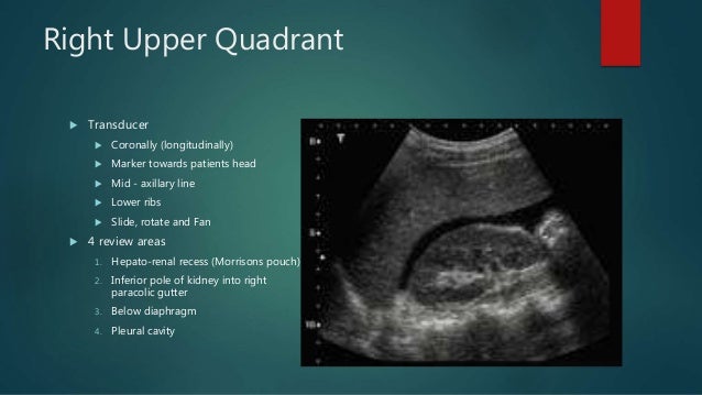
Right Paracolic Gutter Ultrasound

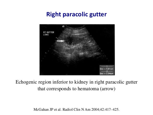
Ultrasonogram Revealed Free Fluid In The Paracolic Gutter Right And Download Scientific Diagram

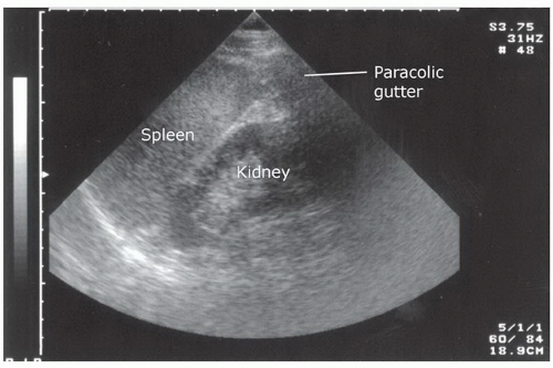
Right Paracolic Gutter Emory School Of Medicine

Ultrasonogram Revealed Free Fluid In The Paracolic Gutter Right And Download Scientific Diagram
Ultrasound Gel
Trauma Radiology Key

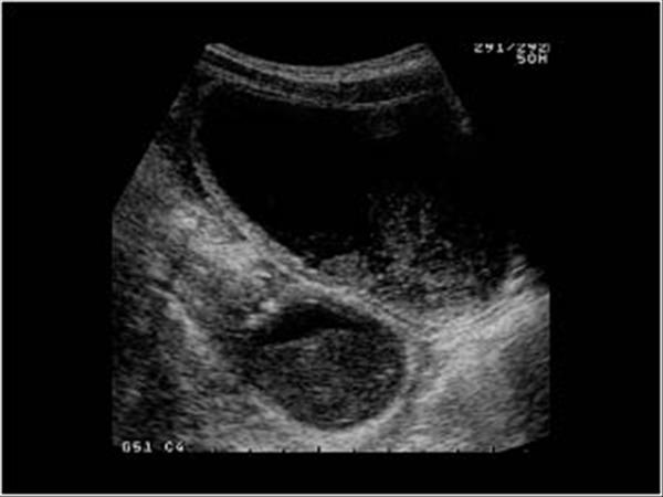
Fluid In The Paracolic Gutter Youtube
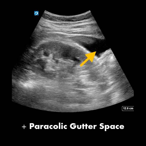
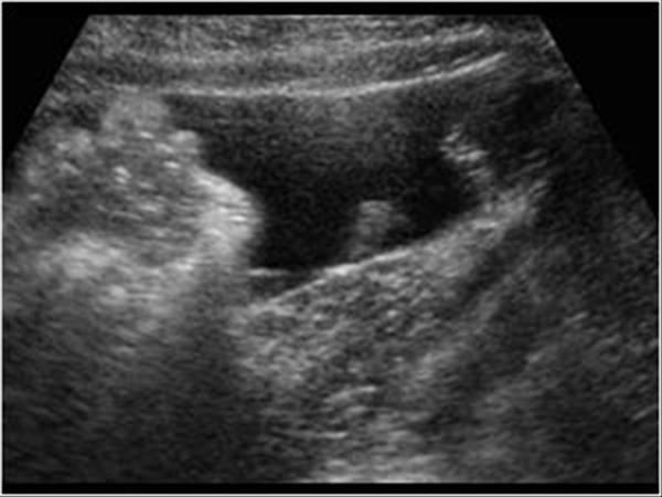
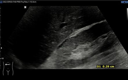
The right lateral paracolic gutter.
Right paracolic gutter ultrasound.

Ultrasound Gel

Positive Right Upper Quadrant Ruq Fast View Showing Superior Download Scientific Diagram

Pancreatic Ascites Radiology Case Radiopaedia Org
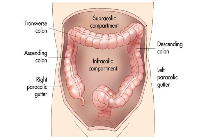
Paracolic Gutter
E Fast Examination
Extended Focus Assessment With Sonography For Trauma

Ultrasonography Showing Fluid Collections Measuring 2 7 Cm In A The Download Scientific Diagram
Https Mafp Org Resource Resmgr Files Sr2019 Handouts Pocus Fastaaa Pdf
Abdomen And Retroperitoneum 1 5 Appendix Case 1 5 3 Complicated Appendicitis Ultrasound Cases

Peritoneal Free Fluid Radiology Key

Endoscopic Ultrasound Of Peritoneal Spaces Abstract Europe Pmc
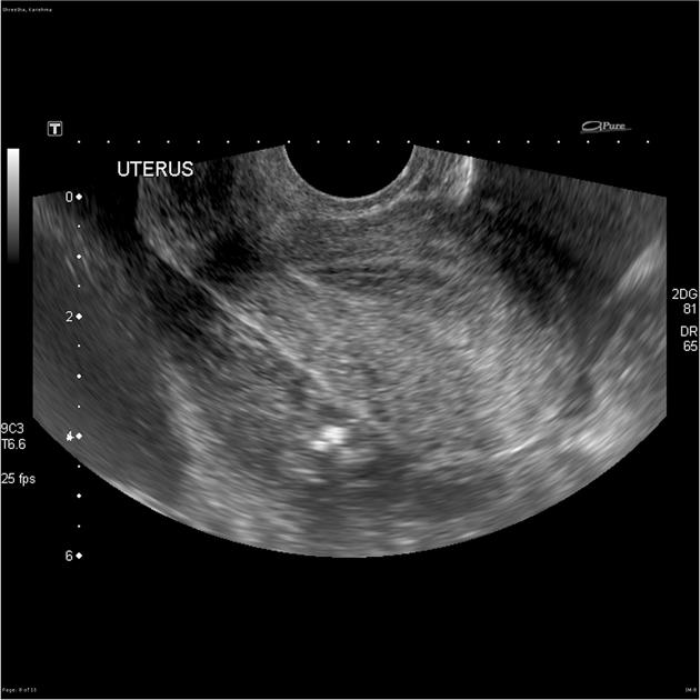
Ruptured Ectopic Pregnancy Radiology Case Radiopaedia Org
Abdomen And Retroperitoneum 1 4 Spleen Case 1 4 7 Trauma Of The Spleen Ultrasound Cases

Fast Exam

Figure 3 From Sonographically Detected Free Appendicolith As A Sign Of Retrocecal Perforated Appendicitis In A 2 Year Old Child Semantic Scholar
Gale Onefile Health And Medicine Document Acute Acalculous Cholecystitis With Empyema Due To Salmonellosis
Posterior Right Subhepatic Space Radiology Reference Article Radiopaedia Org

Splenic Artery Aneurysm Rupture In The Pregnant Patient The Importance Of Utilising Ultrasound To Differentiate The Acute Abdomen From Obstetric Causes Of Abdominal Pain Napier 2019 Sonography Wiley Online Library
Https Encrypted Tbn0 Gstatic Com Images Q Tbn 3aand9gcrbx70kftbqrikhuth2ien3tp Lwqwgoc3vuzrgfdj5xk Zpt5d Usqp Cau
Gale Onefile Health And Medicine Document Whirl Sign And Midgut Volvulus An Unusual Cause Of An Acute Abdomen In An Adult Patient

Perforated Duodenal Ulcer With Valentino Syndrome Radiology Case Radiopaedia Org
Manual Ultrasound

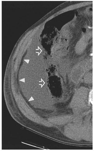
Ct Showing Free Fluid In The Right Paracolic Gutter No Free Air And Download Scientific Diagram

Underused Tool In Radiologist S Toolbox Ultrasound In Unexpected Peptic Ulcer Perforation Semantic Scholar
Source : pinterest.com
